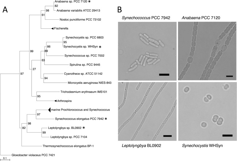
FIG 1.
Diversity of cyanobacterial strains used in this study. (A) Phylogenetic tree of select cyanobacterial species. Species used in this study are marked with asterisks. SH-like branch support values are shown. The evolutionary distance between two sequences is depicted by the sum of the horizontal branches connecting them (scale bar represents 0.1 mutation per position). Gloeobacter violaceus PCC 7421 was used to root the tree. (B) Light micrographs of the cyanobacterial species used in this study. Top left, S. elongatus PCC 7942. Top right, Anabaena sp. strain PCC 7120. Bottom left, Leptolyngbya sp. strain BL0902. Bottom right, Synechocystis sp. strain WHSyn. Scale bars, 5 μm.

