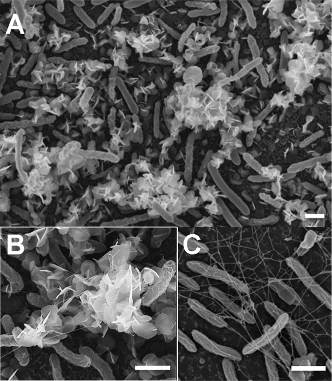
FIG 2.

SEM micrographs of 48-h-old biofilms exposed to 1 mM uranyl acetate for 24 h (A and B) showing the extracellular needle-like, white precipitates of uranium associated with the biofilm microcolonies. (C) Control biofilms not exposed to U are also shown, allowing visualization of the network of extracellular filaments that connect the biofilm cells. Bars, 1 μm.
