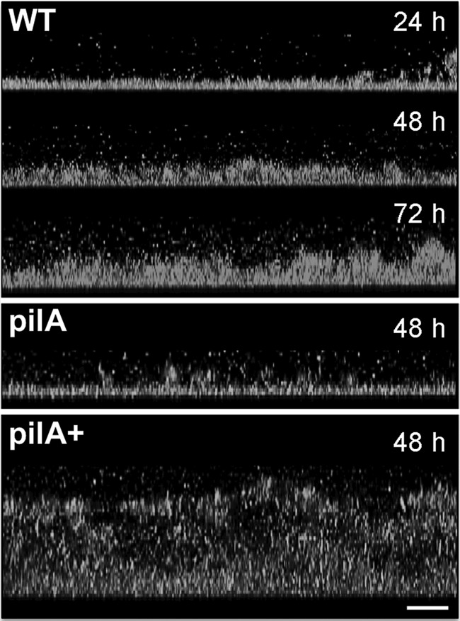
FIG 4.

CLSM micrographs showing side-view projections of WT, pilA, and pilA+ biofilms grown for 24, 48, and/or 72 h. Bar, 20 μm. The biofilm cells were stained with dye solution from the BacLight viability kit. Color top-view projections for these images are shown in Fig. S2 in the supplemental material.
