
FIG 4.
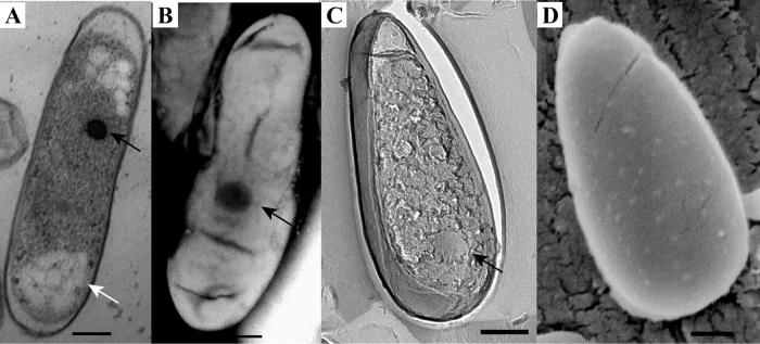
Morphology of the thermophilic verrucomicrobial methanotroph M. fumariolicum SolV as visualized by the use of thin sections of high-pressure-frozen and freeze-substituted cells (A), negative staining (B), FE (C), and cryoSEM (D). The cells are rod shaped with one broader cell pole and contain electron-dense (black arrows) and electron-light (white arrow) particles. Scale bars, 200 nm.