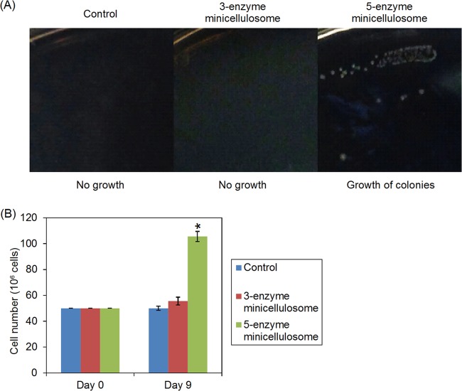
FIG 5.
Growth of minicellulosome-displaying cells. (A) The different strains were grown at 30°C on the same agar plate with 1% PASC as the sole carbon source. Colonies were formed only for the five-enzyme strain after an incubation of 10 days. The colonies were photographed after an incubation of 2 weeks. (B) The different strains were grown in sPASC media containing 1% PASC. Cell numbers were determined by counting with a hemocytometer. Values represent the means of three samples. Error bars denote the standard deviations.

