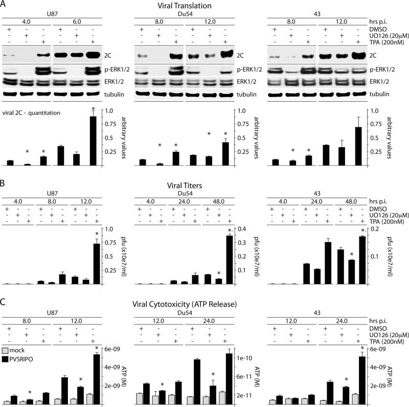
FIG 1.
ERK1/2 signals control PVSRIPO translation, proliferation, and cytotoxicity in GBM cells. (A) U87, Du54, or 43 GBM cells were treated with DMSO (mock), UO126 (20 μM), or TPA (200 nM) (30 min); infected with PVSRIPO; and harvested at the designated intervals. UO126 was maintained after infection; TPA was not. The immunoblots track viral protein (2C); the quantitation represents the average of 3 experiments normalized to the first control value for each series. (B) Supernatants from cells treated and infected as described for panel A were collected to determine viral progeny (PFU); the averages of two experiments are shown. (C) ATP release was measured in supernatants from the experiments in panel B. The ATP concentration was determined using a standard curve; the average of two assays is shown. The error bars represent SEM; the asterisks indicate significant ANOVA-protected t tests.

