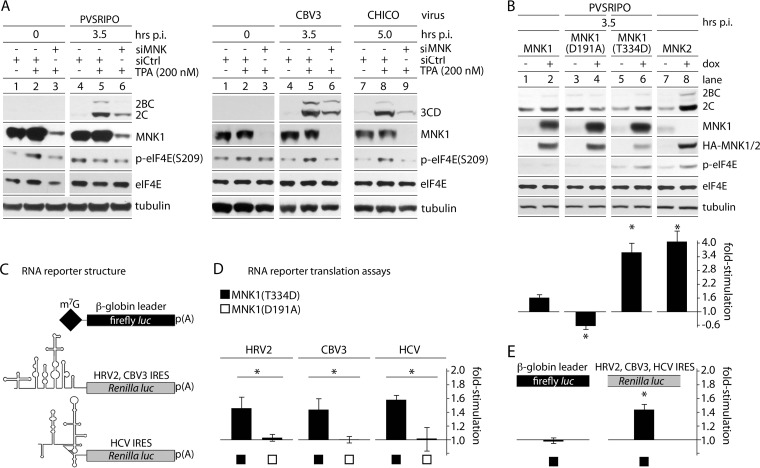
FIG 4.
MNK activity selectively enhances viral IRES-mediated translation. (A) HeLa cells were treated with control (siCtrl) or MNK1-targeting (siMNK) siRNA 24 h prior to TPA/mock stimulation. PVSRIPO (left) or CBV3/CHICO (right) infection and subsequent analyses were carried out as for Fig. 1. The assays were repeated 3 times, and representative series are shown. (B) Cells with Dox-inducible expression of wt MNK1, MNK1(D191A), MNK1(T334D), or MNK2 were mock/Dox induced (12 h) and infected with PVSRIPO. Viral translation was quantitated, and the fold stimulation of viral translation was calculated by dividing the Dox-induced value by the mock-induced value for 3 independent tests. (C) Structure of RNA reporters used (32). (D) Uncapped, in vitro-transcribed rluc RNA reporters driven by the HRV2, CBV3, or HCV 5′ UTR were cotransfected with m7G-capped β-globin leader fluc reporters into Dox-/mock-induced MNK1(T334D)- or MNK1(D191A)-expressing cells (4 h). IRES-driven (rluc) values were divided by β-globin 5′-UTR firefly luciferase values to correct for transfection differences. Dox-induced values were then divided by mock-induced values for each cell line to calculate the fold stimulation of IRES-mediated translation due to MNK1(T334D)/MNK1(D191A) expression. The data represent 3 independent assays done in triplicate for each cell line and 5′ UTR. (E) Pooled β-globin 5′-UTR fluc values and IRES-driven rluc values in MNK1(T334D)-expressing cells. (B, D, and E) The error bars represent SEM, and the asterisks indicate significance (P < 0.05 by Student's t test).

