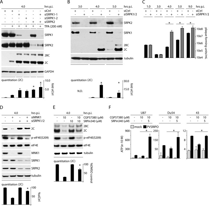
FIG 9.
SRPK depletion enhances PVSRIPO translation and propagation, counters the MNK depletion/inhibition effect, and increases viral translation and cytotoxicity in GBM. (A) HeLa cells were treated with siCtrl (with or without TPA) or siRNA targeting SRPK1 and/or SRPK2 (all plus TPA) prior to infection with PVSRIPO (4 h). The quantitation represents the average viral protein 2C levels normalized for the siCtrl-plus-TPA sample from 2 assays; asterisks denote ANOVA-protected t test. (B) HeLa cells were treated with siCtrl or siRNA targeting SRPK1 and -2 and assessed for viral 2C expression by immunoblotting at 4 and 5 h p.i. Average quantitations from 3 tests, normalized for the control values for each interval, are shown. (C) Viral titers from HeLa cells treated as for panel B and infected at an MOI of 10 were determined for the designated intervals; the average of two experiments is shown, normalizing between experiments using the siCtrl values. (D) HeLa cells were cotransfected with siCtrl or siRNA targeting SRPK1/2 followed by transfection with siCtrl or MNK1 siRNA as shown and infected as for panel B. The quantitation represents the average viral protein 2C levels normalized between experiments by setting siCtrl values to 1. (E) HeLa cells were treated with DMSO (mock), CGP57380 (10 μM), SRPin340 (10 μM), or combined inhibitors coincident with PVSRIPO infection (4 h p.i.). Cells were harvested and analyzed for viral protein by immunoblotting. Quantitation of viral protein 2C is shown for 3 assays normalized by setting the controls (without CGP57380) to 1. (F) U87, Du54, and 43 GBM cells were treated with DMSO, CGP57380, or CGP57380 plus SRPin340 at the concentrations indicated at the time of infection. The supernatants were harvested (12 h p.i.), and the average ATP concentration for 3 (Du54) or 2 (U87 and 43) assays was determined. (B to F) The asterisks denote significant paired Student's t tests; error bars represent SEM.

