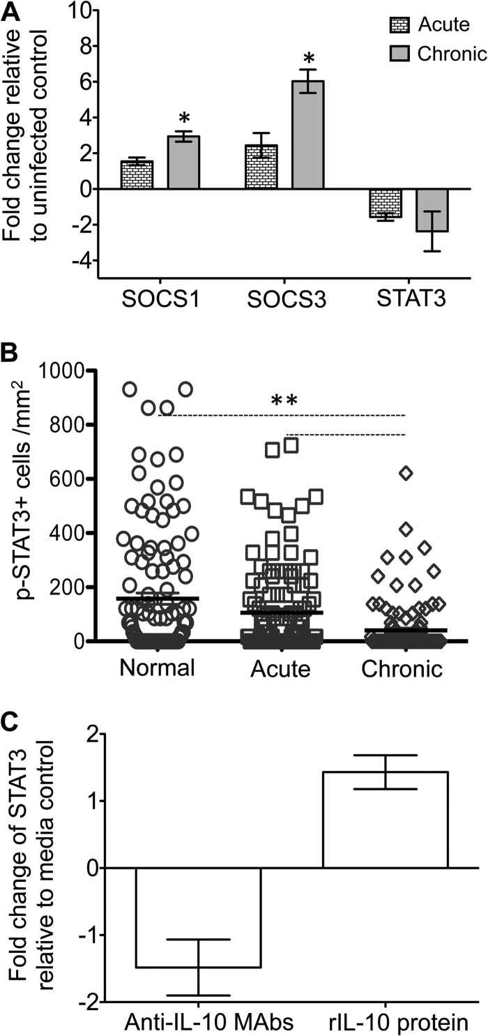
FIG 8.
Expression patterns of SOCS1, SOCS3, STAT3, and pSTAT3 in SIV-infected macaques and colon explant cultures. (A) Increased fold change of SOCS1 and SOCS3 and decreased fold change of STAT3 gene expression were observed in jejunum from acute and chronically SIV-infected rhesus macaques compared to uninfected normal macaques using relative RT-PCR (mean ± the standard error; n = 6). Samples were normalized against 18S rRNA expression. (B) Scatter plots (with means ± the standard errors) of pSTAT3+ cells in uninfected normal macaques and acutely and chronically SIV-infected macaques quantified by immunohistochemistry staining are shown (n = 6; a minimum 20 fields were measured for each sample). (C) The relative fold changes (means ± the standard errors) of STAT3 gene expression are indicated for colon explants in the presence of either anti-IL-10 MAbs or recombinant IL-10 (rIL-10) protein as determined using relative RT-PCR (n = 3). Asterisks indicate significant differences between groups for the specified genes and proteins (*, P ≤ 0.005; **, P < 0.0001).

