
FIG 2.
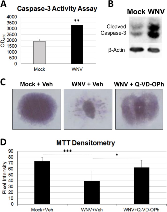
Caspase-dependent apoptotic mechanisms contribute to WNV pathology in SCSCs. (A) A fluorogenic caspase-3 activity assay was used to assess apoptosis in SCSC lysates at 6 dpi by comparison of mock-infected and WNV-infected samples. WNV-infected SCSCs had increased caspase-3 activity compared to mock-infected SCSCs. Asterisks indicate statistically significant differences (**, P = 0.003). OD450, optical density at 450 nm. (B) Western blots were used to determine the amount of cleaved caspase-3 present in lysates prepared from mock-infected and WNV-infected SCSCs at 7 dpi. β-Actin (42 kDa) was present as a loading control. WNV-infected SCSCs have increased cleaved caspase-3 staining at 17 kDa and 19 kDa compared to mock-infected samples. (C) Tissue health was determined with MTT staining in mock-infected and WNV-infected SCSCs at 7 dpi, with significant cell death occurring in WNV-infected samples. Caspase inhibition rescued WNV-infected SCSCs from considerable tissue death. The vehicle (Veh) was DMSO, and the pan-caspase inhibitor was Q-VD-OPh. (D) Densitometry via pixel intensity measurement (with ImageJ software) of MTT-stained images shows significant tissue death from WNV infection and rescue from tissue death with caspase inhibition (for each condition, n = 8). Asterisks indicate statistically significant differences (***, P < 0.001; **, P < 0.01; *, P < 0.05).