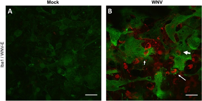
FIG 4.

Microglial phagocytosis in WNV-infected SCSCs. Mock- and WNV-infected samples were collected at 6 dpi and stained for Iba1 (green) and WNV-E (red). (A) Mock-infected SCSCs display low levels of Iba1 staining. (B) WNV-infected SCSCs show increased expression of Iba1 as well as phagocytic functions performed by microglia. Examples of microglial phagocytic activity include partial engulfment of a WNV-E+ cell (short, thin arrow), the formation of a phagosomal compartment with a fully engulfed WNV-E+ cell (long, thin arrow), and a WNV-E+ cell within a microglia cell (short, thick arrow). Bars, 30 μm.
