FIG 5.
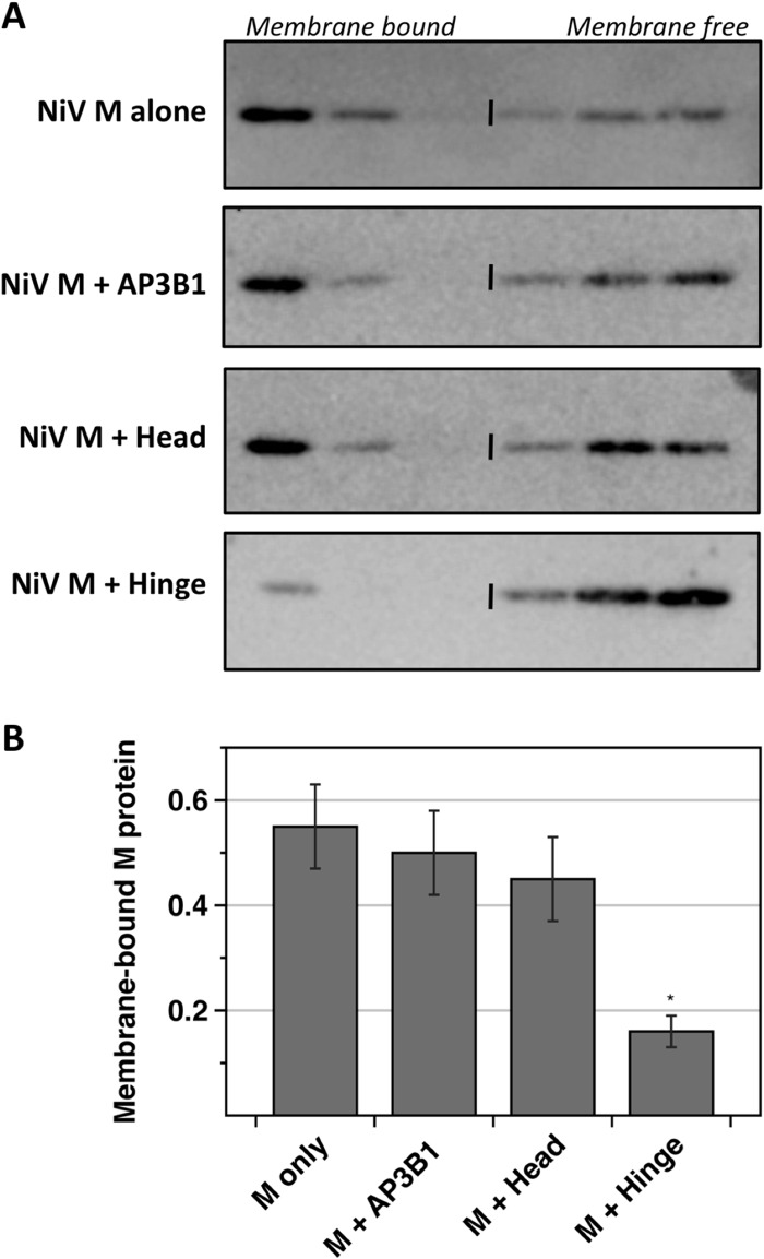
AP3B1 Hinge polypeptide inhibits the membrane-binding ability of Nipah virus M protein. (A) 293T cells were transfected to produce Nipah virus M protein together with the indicated human AP3B1-derived polypeptides. Detergent-free cell lysates were prepared and overlaid with sucrose solutions to form flotation gradients. After ultracentrifugation, samples were collected from the tops of the gradients. Proteins from gradient fractions were resolved by SDS-PAGE, and Nipah virus M protein was detected by immunoblotting using M-specific polyclonal antibody. (B) Three independent experiments were performed as described for panel A. The percentage of M protein that was membrane bound (top three fractions of the gradients) was quantified using a phosphorimager. Results were plotted, with standard deviations indicated by error bars. Differences from the values obtained in the absence of polypeptide coexpression were assessed for statistical significance by using a two-tailed Student t test, and P values of <0.01 are denoted by asterisks.

