
FIG 8.
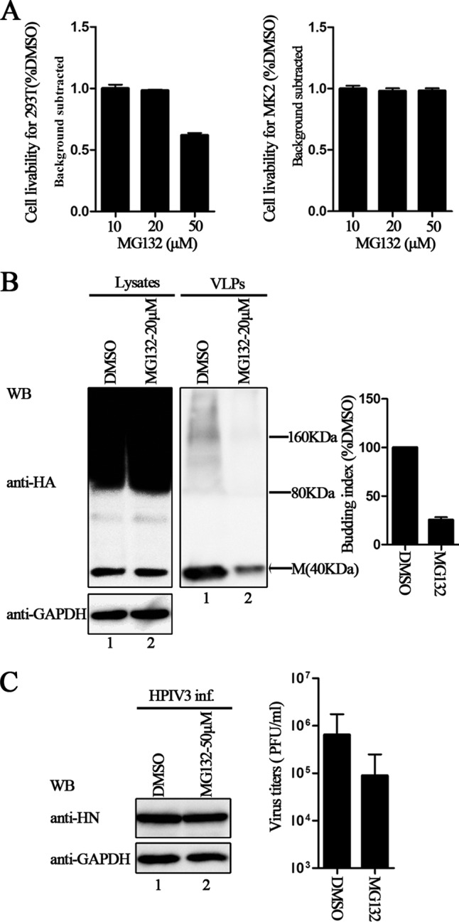
Inhibition effect of proteasome inhibitors on M VLP release and HPIV3 virus titers. (A) Cytotoxicity assays for 293T and MK2 cells were performed as described in Materials and Methods. There was no potential toxicity in either case: when 293T cells were treated with 20 μM MG132 or when MK2 cells were treated with 50 μM MG132. (B) Inhibition of M VLP budding by MG132. 293T cells were transfected with plasmid encoding HA-M and treated with DMSO or 20 μM MG132 for 8 h before harvesting. At 48 h posttransfection, cells and culture medium were harvested, and proteins expressed in both cell lysates and VLPs were immunoblotted with anti-HA Ab. Then, the cell lysate blot was stripped and reprobed with an anti-GAPDH Ab as a loading control. Quantification of the budding index for each group is shown. Standard errors were calculated from three independent experiments. The size markers represent dimer and tetramer of M in VLPs. (C) Inhibition of HPIV3 infection by MG132. MK2 cells infected with HPIV3 were treated with DMSO or 50 μM MG132 for 8 h before harvesting. Cells and supernatants were collected at 36 h postinfection. Cell lysates were immunoblotted with anti-HN Ab and then stripped and reprobed with anti-GAPDH Ab as a loading control, and viral titers of the supernatants were determined by plaque assay as described in Materials and Methods. Standard errors were calculated from three independent experiments.