
FIG 7.
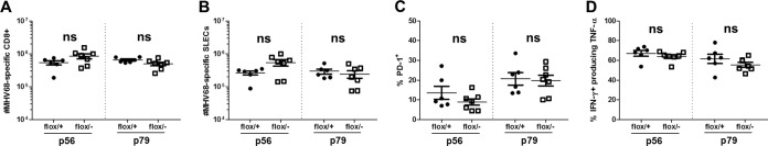
Type I IFN signaling on T cells is dispensable for wild-type CD8+ T cell numbers, effector differentiation, PD-1 expression, and effector cytokine production during MHV68 infection. Lck-Cre, IFNAR1 Fl/+ (flox/+) and Lck-Cre, IFNAR1 Fl/− (flox/−) mice were infected with MHV68 and euthanized at 16 dpi. Splenocytes were labeled with α-CD8, α-CD44, and p56 or p79 tetramers (A), α-CD8, α-KLRG-1, α-CD127, and p56 or p79 tetramers (B), α-CD8, α-PD-1, and p56 or p79 tetramers (C), or α-CD8, α-IFN-γ, and α-TNF-α following stimulation with the indicated peptides (D) and evaluated by flow cytometry. Data points indicate results from individual mice from three independent experiments. Means ± SEM are shown. ns, P > 0.05; *, P ≤ 0.05; **, P ≤ 0.01; ***, P ≤ 0.001 (by paired, two-tailed Student's t test).