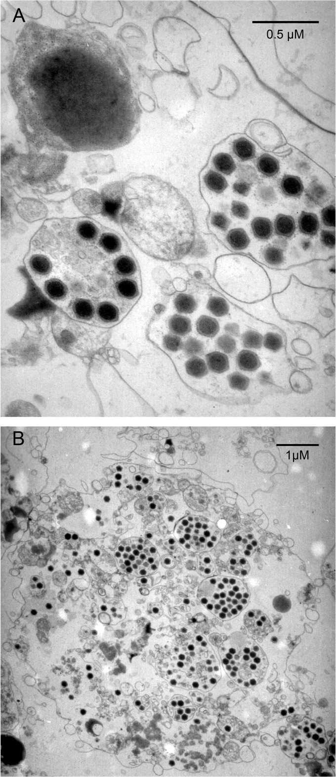
FIG 2.
Electron microscopy images of ultrathin sections of Melbournevirus. (A) Enlarged view of mature Melbournevirus particles in intracytoplasmic vacuoles. (B) Overall view of an infected cell at a late stage of infection, when the cell is about to be lysed. The cell is filled by Melbournevirus mature particles, most of which are in vacuoles. The cytoplasm is disorganized, and the cell organelles are no longer recognizable.

