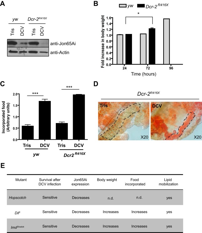
FIG 6.
DCV-induced pathology does not result from activation of immune defense pathways. (A) Jon65Ai is repressed in the gut of Dcr-2 mutant flies. Western blot analysis of proteins extracted from the guts of Tris- and DCV-injected flies 72 h postinfection. (B) Increased body weight in Dcr-2 mutant flies after infection with DCV. The graph represents means ± SD from three independent experiments. All Dcr-2 mutants were dead at 96 h postinfection. (C) Increase of the incorporated food in Dcr-2 mutant flies after infection with DCV. The graph represents means ± SD from two independent experiments. (D) Lipids are mobilized from FB to oenocytes in Dcr-2 mutant flies, as shown by oil red O staining of dissected carcasses from Dcr-2 mutant flies. The dashed lines show the location of the oenocytes. (E) Table showing the development of DCV-induced symptoms in mutants for other antiviral immune pathways, Jak/STAT (Hopscotch), Toll (Dif), and IMD (imd).

