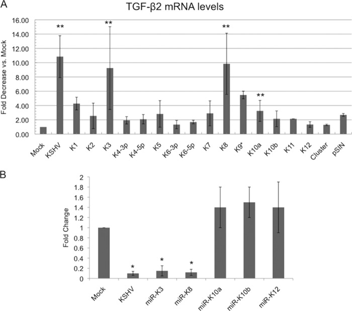
FIG 4.
KSHV miRNAs miR-K3 and miR-K8 downregulate TGF-β2. (A) qRT-PCR analysis of TGF-β2 mRNA from TIME cells that were mock or KSHV infected or transduced with lentivirus expressing individual miRNAs or the control lentiviral vector pSIN. (B) qRT-PCR analysis of TGF-β2 mRNA from TIME cells that were mock or KSHV infected or transduced with a lentivirus expressing KSHV miRNA miR-K3, miR-K8, miR-K10a, miR-K10b, or miR-K12. Cells were harvested, and mRNA was isolated and analyzed by quantitative real-time RT-PCR. Values for samples were normalized to GAPDH values and are reported as fold changes over values for mock-infected cells. Error bars reflect standard errors of the means of data from three independent experiments (*, P < 0.05; **, P < 0.01).

