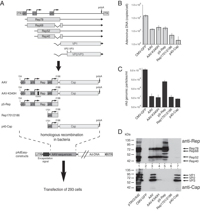
FIG 1.
AAV elements involved in inhibition of adenoviral replication in the context of Ad5/AAV-2 hybrid vectors. (A) Genome organization of AAV-2 and schematic presentation of the AAV-2 sequences introduced into the AdEasy system for generation of recombinant adenoviruses. The viral genome is shown in the upper part of the figure, with the inverted terminal repeats (ITRs), the four Rep proteins (Rep78, Rep68, Rep52, and Rep40), the capsid proteins (VP1 to VP3), and the three promoters, at map units 5, 19, and 40, indicated by different shaded boxes. Right-angled arrows represent the transcription start sites of the promoters, and the vertical arrow indicates the common polyadenylation [poly(A)] site for all transcripts at map position 96. The lower part of the figure shows the AAV-2 sequences cloned into the pAdEasy plasmid by homologous recombination in bacteria. Characteristic nucleotide positions are indicated. (B) Amounts of newly replicated, DpnI-resistant adenoviral DNA obtained 12 days after transfection of PacI-linearized pAdEasy plasmids into HEK-293 cells, determined as numbers of genomic copies per cell by real-time PCR. (C) Amounts of recombinant adenoviral particles obtained in freeze-thaw cell supernatants after transfection of the PacI-linearized pAdEasy plasmids into HEK-293 cells for 12 days and amplification of the primary supernatants in HEK-293 cells for 6 days, determined as numbers of GP/ml of final cell supernatant by real-time PCR. (D) Western blot analysis of whole-cell extracts harvested 2 days after transfection of HEK-293 cells with the indicated pAdEasy-AAV-2 plasmids. Expression of Rep proteins was scored with monoclonal antibody 303.9 (upper panel), and that of capsid proteins was scored with monoclonal antibody B1 (lower panel). Arrows indicate the positions of the different Rep and capsid proteins. HEK-293 cells transfected with infectious AAV-2 plasmid pTAV2-0 and infected with adenovirus type 2 (multiplicity of infection [MOI] = 20) served as a positive control (lane pTAV2/Ad2).

