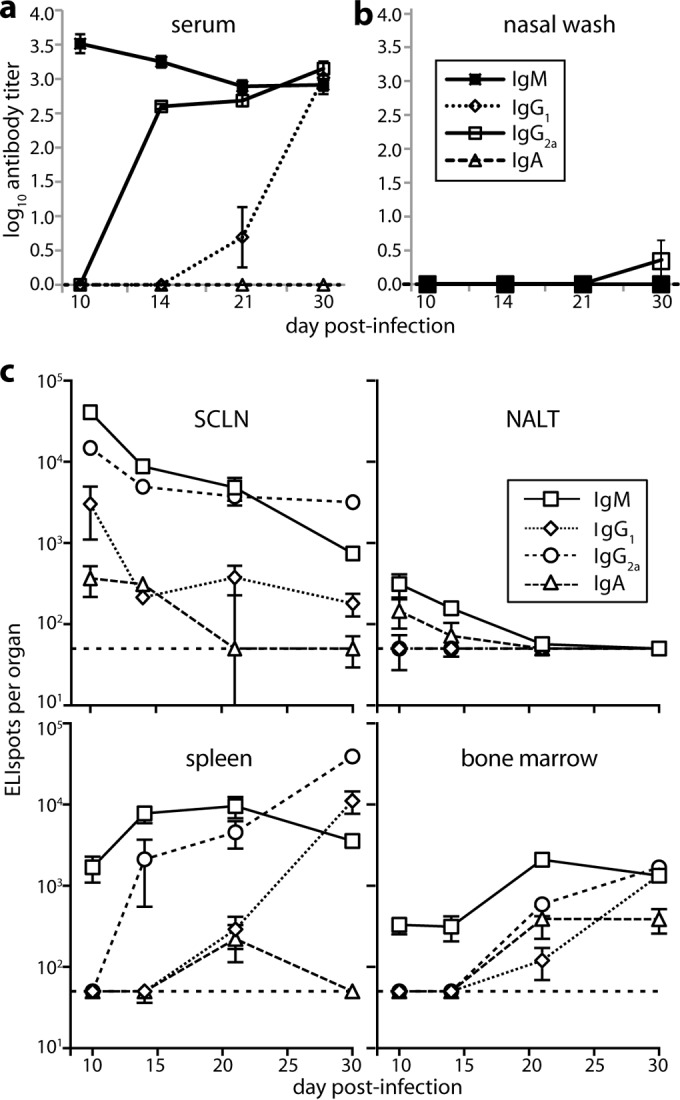
FIG 1.

B cell response to URT MuHV-4 infection. (a) BALB/c mice were infected i.n. with MuHV-4 (105 PFU in 5 μl) and then assayed for virus-specific antibodies by ELISA. Titers are expressed relative to a standard reference of pooled immune sera assayed in parallel. Each point shows the mean ± standard error of the mean (SEM) of 6 mice. The baseline corresponds to the lower limit of assay sensitivity. (b) Mice were infected, and nasal washes were assayed for virus-specific serum antibody by ELISA as for panel a. Each point shows the mean ± SEM of 6 mice. The baseline corresponds to the lower limit of assay sensitivity. (c) Mice were infected as for panel a, and different lymphoid populations were assayed for virus-specific AFCs by ELIspot. Each point shows the mean ± SEM of 6 mice. The horizontal dashed line shows the limit of assay sensitivity (50 AFCs/organ). BM AFC numbers are for only femurs and tibias. Although a larger fraction of the NALT could be assayed due to its small size, it showed more background spots in naive controls. Thus, the limits of assay sensitivity were similar. At day 30 postinfection, SCLN (P < 0.01), spleen (P < 0.005), and BM (P < 0.001) contained significantly fewer IgA+ AFCs than IgM+, IgG1+, or IgG2a+ AFCs.
