FIG 5.
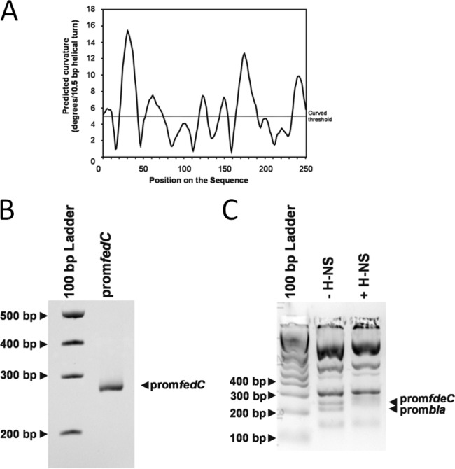
H-NS binds to the promoter region of fdeC. (A) In silico curvature propensity plot showing predicted regions of curved DNA in the 250-bp PCR fragment containing the fdeC promoter. (B) Electrophoretic mobility of the 250-bp PCR fragment containing the fdeC promoter. The migration of this band is slightly retarded in the gel, suggesting that the DNA is curved. (C) Electrophoretic band shift of the 250-bp PCR fragment containing the fdeC promoter and the bla promoter from TaqI-SspI-digested pBR322 DNA in the absence or presence of 4 μM H-NS. The migration of pBR322 fragments not containing the bla promoter was not altered by H-NS. The image depicts a gel representative of three independent experiments.

