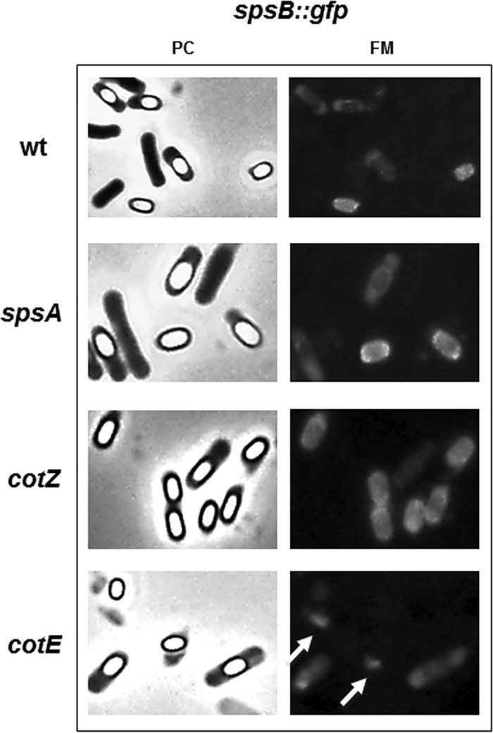
FIG 3.

Fluorescence microscopy analysis of strains carrying GFP fused to SpsB. A representative microscopy field is shown by phase contrast (PC) and fluorescence (FM) microscopy. Fluorescence signals are diffuse in sporulating cells and localized around mature spores of the wild-type strain and spsA and cotZ null mutants. In a cotE null mutant, SpsB-GFP fluorescence is diffuse in sporulating cells but is not found around mature spores. Fluorescent spots observed outside free spores of the cotE mutant (white arrows) but not observed with the other strains are fluorescent material most probably detached from spores.
