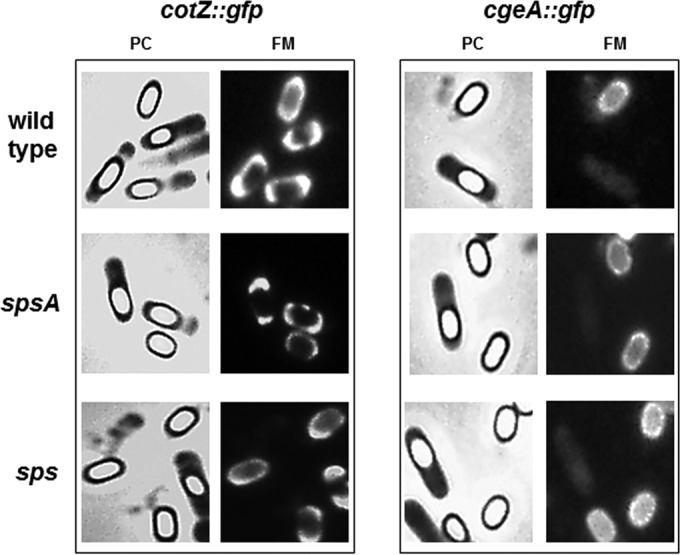
FIG 4.

Fluorescence microscopy analysis of strains carrying GFP fused to CotZ or CgeA. A representative microscopy field is shown by phase contrast (PC) and fluorescence (FM) microscopy. Fluorescence signals are found around mature and forming spores of the wild type and both sps mutants.
