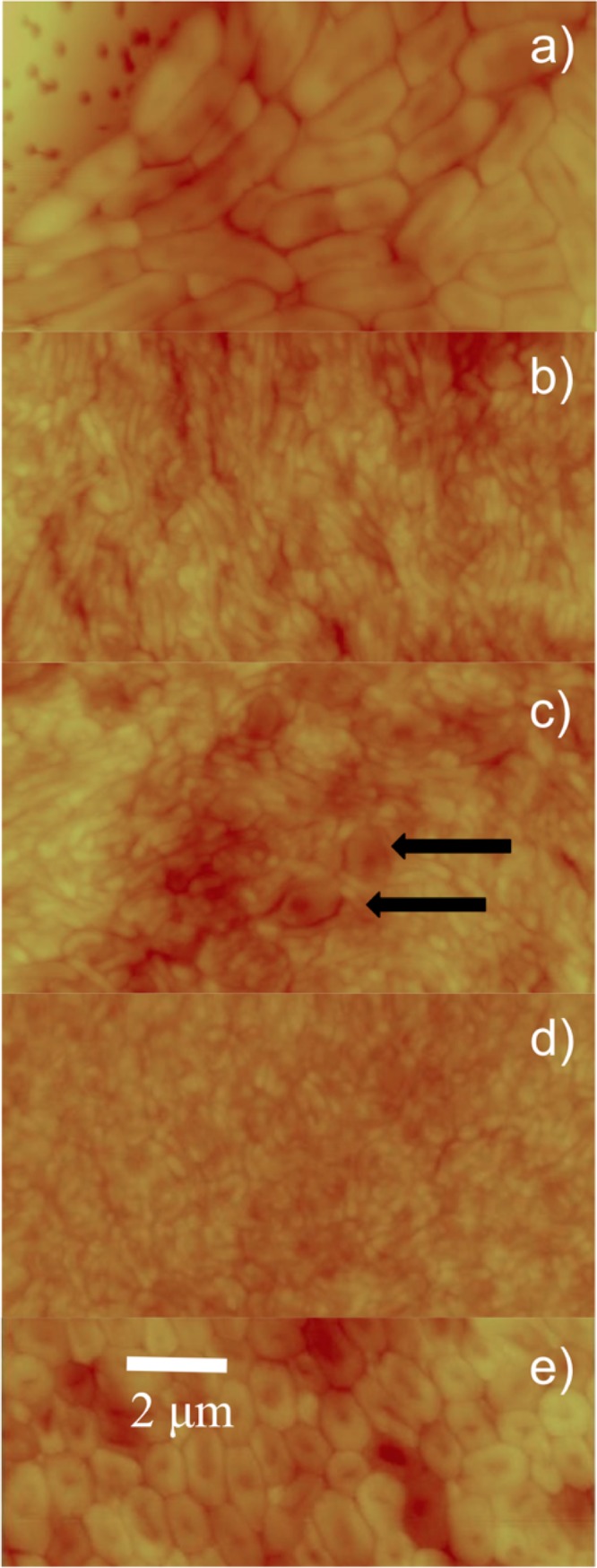
FIG 4.

AFM images of the mature film. Contact AFM images (constant-force mode) in air were collected from the top layer of cells from a mature, spatially resolved film. The film was sampled at the outermost edge (a), the area where inner and outer regions meet (b and c), and the inner region (d). A control image of a film of E. coli ML-35 is presented in panel e. The scale bar in panel e applies to all images; panels a to d have a z scale of 1 μm, while panel e has a 0.5-μm z scale. In panel a, the filter holes of the membrane are visible. Black arrows in panel c indicate bdelloplasts.
