
FIG 2.
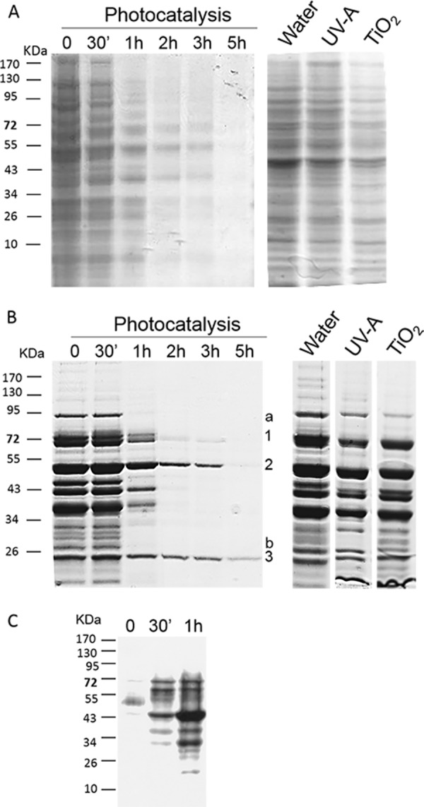
Fate of extracted proteins during photocatalytic treatment. S. cerevisiae cells were incubated in the dark in the presence of TiO2 for 30 min, and an aliquot was harvested (lane 0). The suspension was then illuminated by UV-A, and samples were harvested after 30 min and 1, 2, 3, and 5 h of photocatalytic treatment. Insoluble proteins, i.e., parietal and membrane proteins (A), and intracellular soluble proteins (B) were extracted from each sample and analyzed by SDS-PAGE and Coomassie blue staining. As controls, the results for insoluble and soluble proteins extracted from S. cerevisiae cells incubated for 5 h in the presence of water, UV-A, or TiO2 are presented (right panels). (C) Carbonylated proteins were detected by Western blotting with an anti-DNP antibody in samples collected after 0 min, 30 min, and 1 h of photocatalytic treatment. In panel B, the numbers (1, 2, and 3) indicate slowly disappearing sequenced proteins, while letters (a and b) indicate rapidly disappearing sequenced proteins.