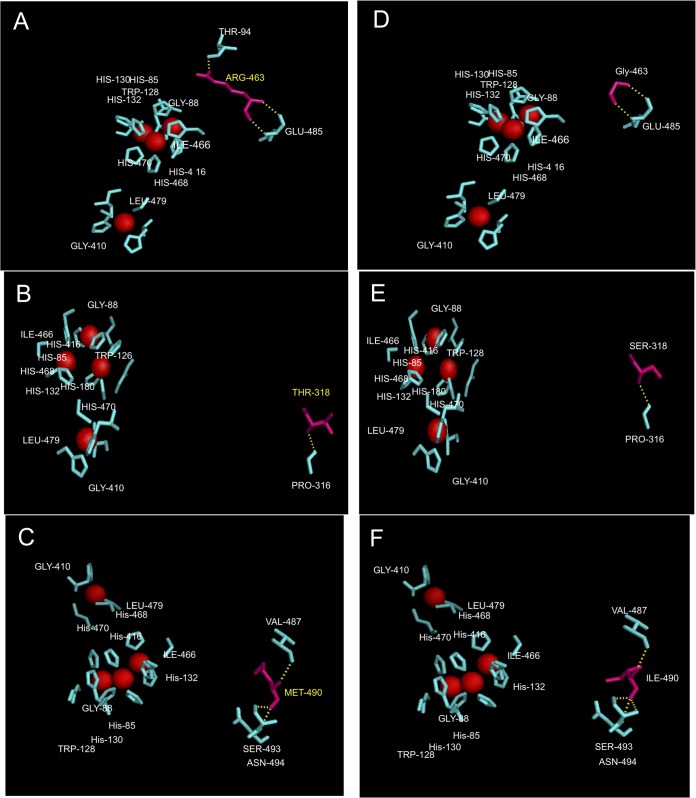
FIG 6.
Details of the point mutations observed in Lcc-35 (G463R) (A), Lcc-61 (S318T) (B), and Lcc-62 (I490M) (C) modeled in the WtLcc structure. Corresponding positions of the native amino acids at these locations are shown in panels D, E, and F, respectively. Red spheres represent the Cu atoms. Hydrogen bonding before and after mutation is shown as yellow dashes.

