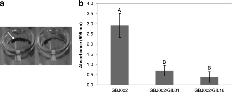
FIG 4.
Biofilm formation of B. thuringiensis serovar israelensis strain GBJ002 and derivative lysogenic strains GBJ002/GIL01 and GBJ002/GIL16. (a) Biofilm formation at the air-liquid interface by nonlysogenic strain GBJ002. Arrow, the pellicle ring stained by crystal violet. (b) Biofilm quantification determined by the crystal violet staining method using measurement of the absorbance at 595 nm. Each plotted datum represents the average of five replicate wells. The error bars indicate the standard deviation values. Means with different letters (A, B) indicate significant differences (Tukey's test, P < 0.05).

