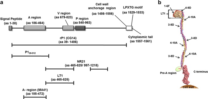
FIG 1.
Schematic representation of functional and structural domains of the S. mutans P1 protein. (a) Linear representation of the protein sequence with the indicated functional/structural domains. The corresponding amino acid positions are shown in parentheses. Bars placed below the schematic indicate the recombinant proteins used in the present study: CG14 (recombinant full-length P1 sequence [rP1]), P139–512, MA41 (A region), LT1 (globular segment intervening between the A and P regions), and NR21 (fusion polypeptide lacking the P region). (b) Tertiary structural model of the S. mutans P1 protein. The location of P139–512 is marked by a red dotted line. The location of the LT1 polypeptide is indicated by a bracket. The approximate positions of experimentally determined epitopes recognized by the P1-specific MAbs (1-6F, 4-9D, 4-10A, and 3-8D) are indicated by arrows. The crystal structure of the N terminus has not yet been determined and is represented as an ellipse. It is known to interact with the C terminus to form a stable complex.

