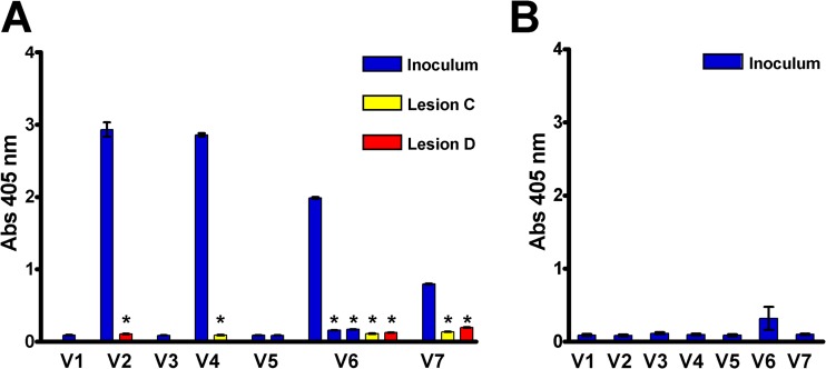
FIG 3.
TprK variants found in secondary lesions escape antibody binding. (A) Antibody binding (ELISA) of antiserum (collected at the time of development of secondary lesions [60 days postinfection]) from rabbit 7912 against synthetic peptides representing V region sequences identified in the inoculum (blue bars) and in two secondary lesions (red and yellow bars) that arose in the rabbit. Antibody reactivity is seen to the predominant sequences of the inoculum V2, V4, V6, and V7 regions, but significantly lower reactivity was detected to the V region sequences found in the secondary lesions. Note also that there is significantly lower antibody reactivity to the two minority V6 sequences that were present in the inoculum (also in blue). Sera from all rabbits with secondary syphilis were similarly tested and showed the same pattern of reactivity to the majority inoculum sequences, but not to the minor inoculum sequences or the variant secondary-lesion sequences. (B) Mean (±standard error [SE]) ELISA results for antisera (collected at the time of lesion appearance [mean, 22 days postinfection]) from rabbits with disseminated primary syphilis (n = 5 rabbits) showing lack of antibody reactivity directed to the V region sequences from the inoculum. *, two-tailed, paired Student t test; P < 0.001 compared to antibody binding to the peptide representing the majority sequences identified in the inoculum. Abs 405 nm, absorbance at 405 nm.

