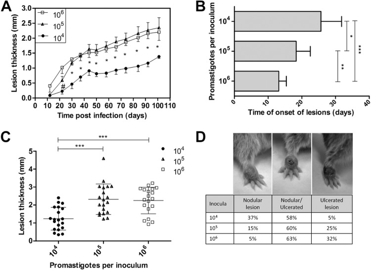
FIG 1.
Evaluation of the golden hamster skin lesions that were infected with Leishmania (Viannia) braziliensis at three different inoculum concentrations (104, 105, and 106 parasites). The lesion sizes were measured by evaluating the dorsal-ventral thickness differences between the infected and uninfected paws. These data are expressed in millimeters. (A) Skin lesion development kinetics through the 105 days following the infections. The graph represents four independent experiments (median and standard error; n = 59 animals). (B) The onset of clinical skin lesions for each inoculum concentration, given in days postinfection (mean ± SD); (C) final skin lesion measurements 105 days after infection (mean ± SD; n = 59 animals; P < 0.0001); (D) final lesion clinical aspect images 105 days after infection. Left, a nodular lesion; middle, a nodular and an ulcerated lesion; right, an ulcerated lesion. #, a significant difference was observed only between the 104 and 106 parasite-infected groups (P < 0.05). *, P < 0.05; **, P < 0.01; ***, P < 0.001.

