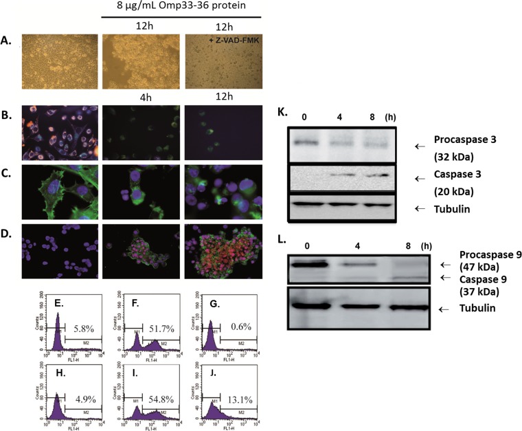
FIG 2.
(A) Morphological changes indicative of apoptosis by bright-field microscopy. The changes in HEp-2 cells incubated with 8 μg/ml of Omp33-36 for 12 h (center and right) and in the absence of this protein (left) are shown. The cells were pretreated for 1 h with 5 μM the caspase inhibitor Z-VAD-FMK (right). Magnifications, ×20. (B to D) Apoptotic features analyzed by fluorescence microscopy. (B) Untreated cells accumulated JC-1 (red) in polarized mitochondria (left). In cells incubated with Omp33-36 for 4 h (center) or 12 h (right), the green monomeric form of JC-1 accumulated throughout the cytoplasm (both images, center and right). Magnifications, ×40. (C) Costaining of the cytoskeleton with phalloidin and of nuclear DNA with DAPI. Round nuclei and a structured cytoskeleton are evident in untreated cells (left), whereas cells incubated with Omp33-36 for 4 h (center) or 12 h (right) show a collapsed cytoskeleton and apoptotic bodies. Magnifications, ×100. (D) Treated cells were stained with DAPI, PI, and annexin V, and untreated cells were treated with DAPI only. Cells incubated with Omp33-36 for 4 h (center) and 12 h (right) were stained with annexin V and in some cases with PI, which specifically stains fragmented DNA. Magnifications, ×40. The images are representative of those from three independent experiments, in which at least 50 cells were scored each time. (E to J) Flow cytometric analysis of apoptosis by staining by use of an APO-BrdU kit (TUNEL assay). HEp-2 cells (E to G) and HeLa cells (H to J) were seeded at a density of 5 × 10⁵ cells and were then either challenged with 8 μg Omp33-36/ml (F, G, I, J) shortly thereafter or left untreated (E, H). To block Omp33-36, the cells were incubated for 1 h at 4°C with excess anti-Omp33-36 antibody, which was added to the cultures together with Omp33-36 (G, J). After 12 h, the cells were harvested and analyzed for apoptosis, as described in Materials and Methods. (K, L) Caspase cleavage during Omp33-36-induced apoptosis. HEp-2 cells were challenged at different times with 8 μg Omp33-36/ml. Cell lysates were resolved by SDS-PAGE on 12% gels and immunoblotted with specific antibodies. Tubulin served as a loading control. (K) Lysates prepared from HEp-2 cells exposed to 8 μg/ml of Omp33-36 for 4, 8, and 12 h were blotted against procaspase 3 and caspase 3. (L) Lysates prepared from HEp-2 cells exposed to 8 μg/ml of Omp33-36 for 4 and 8 h were blotted against procaspase 9 and caspase 9.

