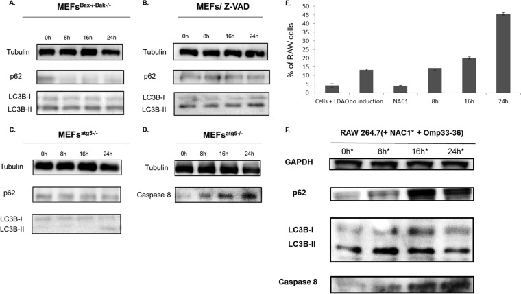
FIG 6.
(A, B) Activation of the autophagy mechanism occurs without development of apoptosis. Western blotting was used to study the autophagy markers p62 and LC3B in cells defective for apoptosis (bax−/− bak−/− MEFs [MEFsBax−/−Bak−/−] and wt MEFs pretreated with 5 μM Z-VAD [MEFs/Z-VAD]). The cells were incubated with 8 μg/ml of Omp33-36 for 0, 8, 16, and 24 h. (C, D) Apoptosis occurs without autophagy. Western blotting was used to analyze the autophagy markers (p62 and LC3B) and apoptosis antibodies (caspase 8) in defective autophagic cells (atg5−/− MEFs). Cells were also incubated with 8 μg/ml of Omp33-36 for 0, 8, 16, and 24 h. (E) Time course of ROS production by cells subjected to oxidative stress (indicated as the percentage of RAW 264.7 cells) after incubation with Omp33-36 (8, 16, and 24 h). Negative controls: cells + LDAO, Omp33-36 buffer solution; no induction, cells alone; and NAC1, cells plus 20 mM NAC1. All negative-control experiments were done at 24 h. There was a significant increase (P < 0.001, Mann-Whitney U test) in ROS production over time in the presence of Omp33-36. (F) Inhibition of ROS response as consequence of apoptosis and blockage of autophagy. An immunoblot with anti-LC3B, anti-p62, and anti-caspase 8 antibodies in RAW 264.7 cells treated for 30 min with 20 mM NAC1 and 8 μg Omp33-36/ml at 0, 8, 16, and 24 h is shown.

