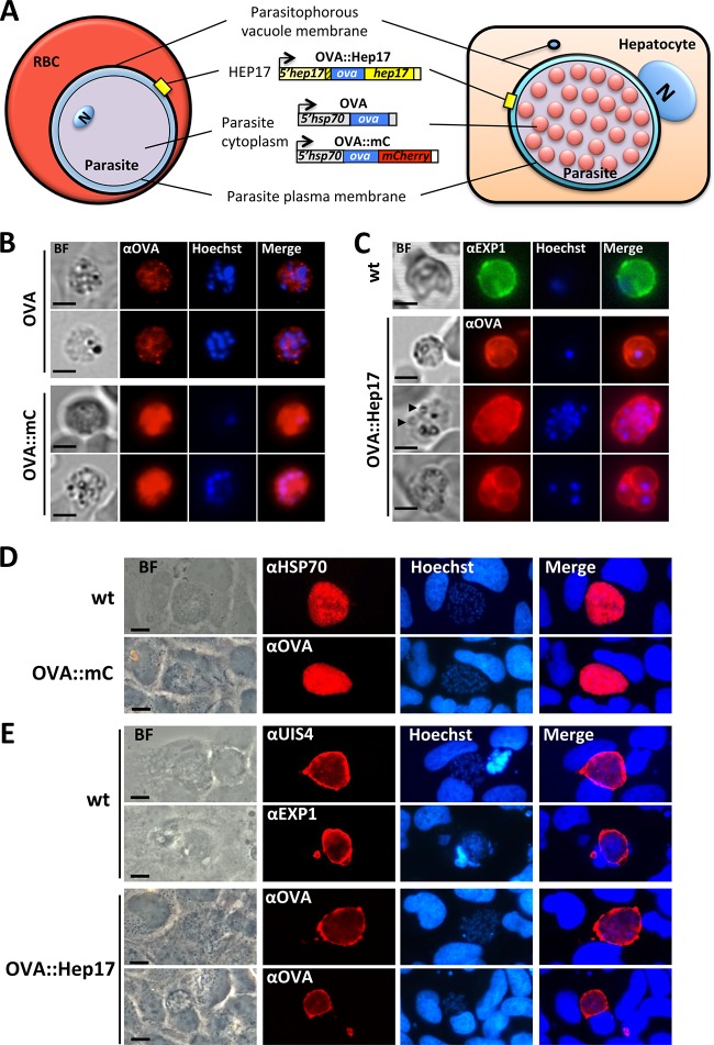
FIG 2.
Subcellular locations of OVA in different transgenic P. berghei ANKA lines. Transgenic P. berghei parasites expressed full-length OVA either under the control of the constitutive hsp70 promoter, unconjugated or fused to mCherry (OVA and OVA::mC, respectively), or under the control of the hep17 promoter, where OVA was fused to the PVM protein HEP17/EXP1 (OVA::Hep17). (A) Schematics showing the expected localization of OVA in the 3 transgenic lines during blood-stage (RBC) and liver-stage (hepatocyte) development. N, nucleus (parasite nucleus in the RBC and hepatocyte nucleus in the liver cell). (B) Immunofluorescence analysis of blood-stage (schizont) parasites stained with anti-OVA antibodies (red) shows a low-level cytoplasmic expression in OVA parasites, whereas in OVA::mC parasites, a very strong cytoplasmic expression of OVA is observed. Trophozoites and schizonts are shown (single and multinucleated cells, respectively). Nuclei were stained with Hoechst 33342 (blue). (C) In OVA::Hep17 parasites, anti-OVA staining reveals a circumferential pattern at the periphery of the parasites, indicative of localization at the PVM, which is very similar to the PVM pattern observed for wild-type (wt) blood-stage parasites by use of antibodies against HEP17/EXP1. A trophozoite (single nuclei) and a fully segmented schizont (with individual merozoites indicated by arrowheads) are shown surrounded by OVA, with a pattern indicative of the PVM. A multiply infected erythrocyte (bottom panels) is also shown, with each individual parasite inside the red blood cell also surrounded by an OVA-stained structure, again indicating the PVM signal. (D) Immunofluorescence analysis of cultured Huh7 hepatocytes infected with OVA::mC (at 40 h post-sporozoite infection [hpi]), using anti-OVA antibodies (red), reveals a cytoplasmic expression for OVA. Intracellular wild-type parasites (at 40 hpi) stained with anti-HSP70 antibody show a similar cytoplasmic localization pattern. (E) Immunofluorescence analysis of cultured Huh7 hepatocytes infected with OVA::Hep17 parasites at 48 hpi, using anti-OVA antibodies (red), reveals a clear circumferential staining around the periphery of the parasites. This staining pattern is very similar to the PVM pattern observed for wild-type liver-stage parasites (also at 48 hpi) by using antibodies against the PVM markers EXP1 and UIS4 (PBANKA_050120). Nuclei were stained with Hoechst 33342 (blue). BF, bright field. Bars = 5 μm (B and C) and 10 μm (D and E).

