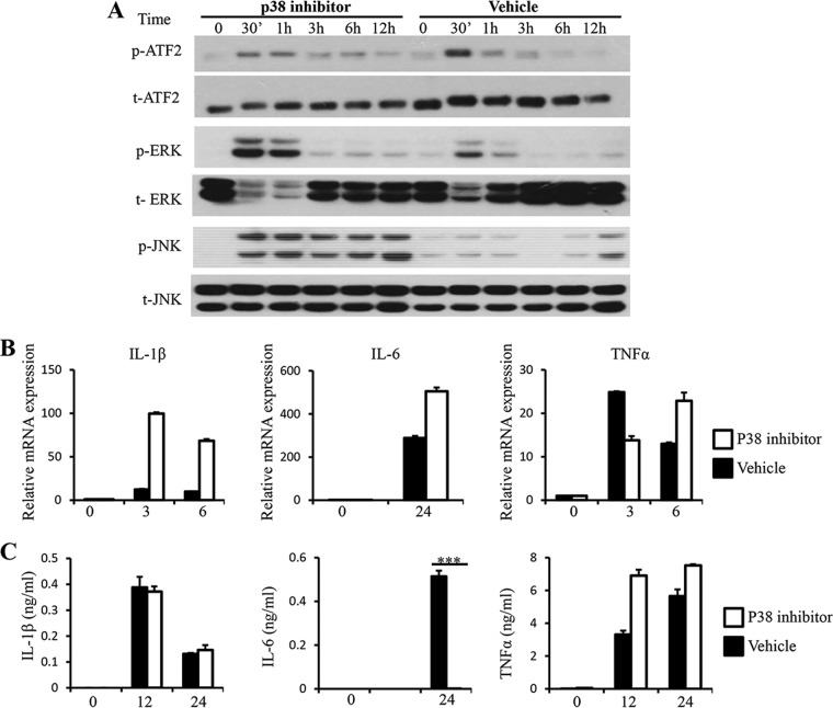
FIG 5.
Inhibition of p38 leads to increased phosphorylation of ERK and JNK and reduced secretion of IL-6. RAW264.7 cells were pretreated with either 20 μM SB203580 or DMSO vehicle dissolved in RPMI complete medium for 1 hour prior to stimulation with Blastocystis ST-7 at an MOI of 10. (A) Representative Western blots for activation of ATF2, ERK, and JNK. Cells were lysed and processed by SDS-PAGE. Membranes were then blotted with phospho-ERK, ERK, phospho-JNK, JNK, phospho-ATF2, and ATF2 antibodies. Each blot is representative of three separate experiments. (B) mRNA levels of TNF-α, IL-1β, IL-6, and GAPDH were measured by qRT-PCR. The values are expressed as mean relative transcript levels normalized to GAPDH and are expressed relative to the 0-h time point, which is set at 1. (C) Concentrations of IL-6, TNF-α and IL-1β in the culture supernatants were measured by ELISA. Each value represents the mean and standard deviation from at least 3 individual experiments. ***, P < 0.005 (representing a statistically significant difference between inhibitor and vehicle treatments).

