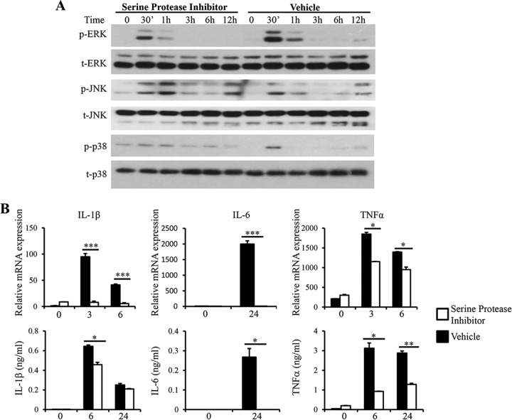
FIG 6.
Serine protease inhibition leads to decreased ERK activation and proinflammatory cytokine expression. ST-7 (B) lysates were pretreated with 500 μM serine protease inhibitor, AEBSF, for 1 hour before coincubation with RAW264.7 cells at 37°C. (A) Representative Western blots for activation of MAPKs. Cells were lysed, and cell extracts were analyzed by Western blotting using phospho-ERK, total ERK, phospho-JNK, total JNK, phospho-p38, and total p38 antibodies. (B) mRNA levels of TNF-α, IL-1β, IL-6, and GAPDH were measured by qRT-PCR. The values are expressed as mean relative transcript levels normalized to GAPDH and are expressed relative to the 0-h time point, which is set at 1. (C) Concentrations of IL-6, TNF-α, and IL-1β in the supernatants were measured by ELISA. Each value represents the mean and standard deviation from at least 3 individual experiments. *, P < 0.05; **, P < 0.01; ***, P < 0.005 (representing a statistically significant difference between inhibitor and vehicle treatments).

