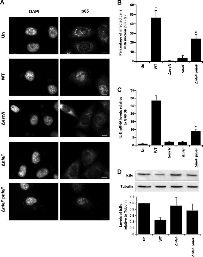
FIG 6.
Early activation of p65 nuclear translocation in infection is dependent on NleF. (A) Immunofluorescence images of p65 localization using anti-N-terminal p65 in HeLa cells infected with a WT, ΔescN, ΔnleF, or ΔnleF pnleF EPEC strain for 1.5 h. Cell nuclei were visualized with DAPI. Bars, 10 μm. (B) Counts were taken in 8 separate fields of view for each condition of cells exhibiting nuclear p65. A significant difference in p65 nuclear localization was seen between uninfected cells (Un) and WT-infected cells and between uninfected cells and cells infected with the ΔnleF pnleF strain. Results are averages for three independent biological repeats. Asterisks indicate significant differences (P < 0.05) from results for uninfected cells. (C) Representative IL-8 expression levels of HeLa cells infected with a WT, ΔescN, ΔnleF, or ΔnleF pnleF EPEC strain for 1.5 h. IL-8 mRNA levels were analyzed by RT-qPCR. IL-8 expression levels were significantly higher for cells infected with the WT or ΔnleF pnleF strain than for cells infected with the ΔescN strain. Results are averages for at least two independent biological repeats. Asterisks indicate significant differences (P < 0.05) from results with the ΔescN strain. (D) Levels of IκBα were analyzed by Western blotting 1.5 h after infection with the EPEC WT, ΔnleF, or ΔnleF pnleF strain. Cells infected with the WT or ΔnleF pnleF strain had lower levels of IκBα than uninfected cells and cells infected with the ΔnleF strain, relative to tubulin levels. Results are representative of those for two independent biological repeats.

