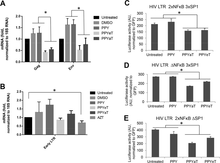
FIG 8.
Effects of PPY-based iron chelators on HIV-1 mRNA expression, HIV-1 reverse transcription, and basal HIV-1 transcription. (A and B) THP-1 cells were left uninfected or infected with HIV-1 Luc and then left untreated or treated with DMSO, AZT, 1 μM PPY (control compound), 1 μM PPYeT, or 1 μM PPYaT, as indicated, for 48 h (A) or 6 h (B). RNA (A) or DNA (B) was extracted. RNA was reverse transcribed and analyzed with primers for the HIV-1 gag and env genes by real-time PCR on a Roche 4800 machine, using 18S RNA as a reference. DNA was analyzed by real-time PCR on a Roche 4800 machine, using primers for early and late LTRs and with the β-globin gene as a reference. (C and D) Effects of PPY-based iron chelators on basal HIV-1 transcription. 293T cells were transiently transfected with vectors expressing the HIV LTR followed by the luciferase reporter gene (WT HIV LTR 2×NFκB 3×SP1 [C], HIV LTR ΔNFκB 3×SP1 [D], or HIV LTR 2×NFκB ΔSP1 [E]; see Materials and Methods for details on the vectors). For normalization, the cells were also cotransfected with a GFP-expressing vector. At 24 h posttransfection, the cells were treated with 10 μM PPY-based iron chelators or the PPY control for 24 h. The cells were then lysed, and luciferase activity was measured. GFP fluorescence was measured in parallel and used for normalization. *, P ≤ 0.01.

