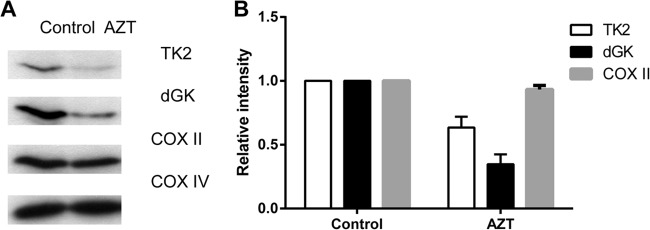
FIG 1.

TK2 and dGK protein levels in cells treated with AZT. (A) U2OS cells were incubated in the presence or absence of 20 μM AZT for 3 days, and then mitochondria were isolated and used to determine the levels of TK2 and dGK by Western blot analysis using the corresponding antibodies. (B) The band intensities were quantified and are shown as TK2, dGK, and COX II levels relative to the controls after normalization to the level of COX IV.
