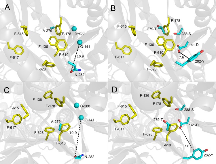
FIG 1.
Positions of the most frequently selected mutations from in vitro random mutagenesis of the periplasmic domain of AcrB (drug/NMP selection). Nonmutated (A and C) and mutated (B and D) side chains are shown as cyan sticks (glycines as cyan spheres), and distal binding pocket phenylalanine side chains are shown as yellow sticks. A side view from the periplasmic outer face (A and B) and a top view from the TolC docking domain (C and D) of the binding state AcrB protomer (PDB code 2HRT; www.rcsb.org/pdb) are shown. Dotted lines show distance (Å) between side chains of the double mutation. Images were created using PyMOL (www.pymol.org).

