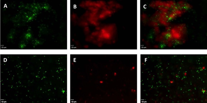
FIG 2.
Viable bacteria are present within P. aeruginosa aggregates formed in the presence of lysed neutrophils. Strain PAO1-GFP was combined with neutrophil lysates for 48 h. Samples were stained with propidium iodide to distinguish nonviable bacteria. (A and D) Viable bacteria (green) occur in clusters within a larger aggregate (A) but are dispersed by DNase treatment (D). (B and E) Nonviable bacteria and extracellular DNA identified by propidium iodide (red) in aggregates (B); treatment with DNase reduces propidium iodide staining (E). (C and F) Overlay of red fluorescent and green fluorescence bacteria. (D to F) Samples treated with DNase, resulting in near complete disruption of the aggregates. Viewed with a 40× objective; length of size bar, 10 μm. Representative images from 9 independent experiments.

