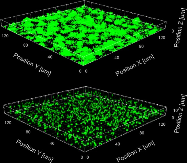
FIG 5.
Three-dimensional confocal images of 1-day-old miniTn7-gfp-tagged P. aeruginosa PAO1 slide biofilms with either ABTGC medium (control, top panel) or 500 μM iberin (bottom panel). Images were obtained using confocal microscopy at ×63 magnification (Zeiss LSM 780 confocal system), and analyzed using the Imaris software package (Bitplane AG). Only a representative image of three replicates is shown.

