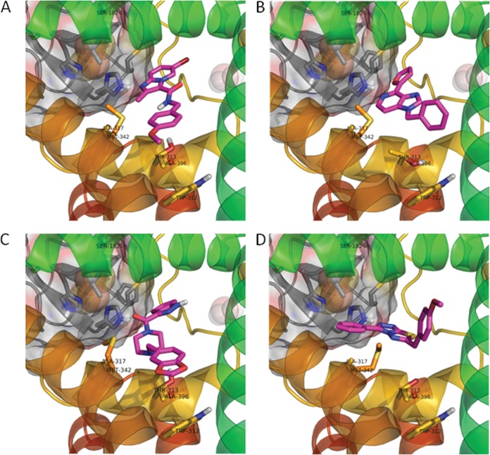
FIG 2.
Modeling of the imidazopyridine binding pocket of QcrB showing docking poses for the two scaffolds. (A) Docking of imidazo[1,2-α]pyridine (compound 2) in the QcrB homology model; (B) docking of imidazo[4,5-c]pyridine (compound 1) in QcrB; (C) docking of compound 3; (D) docking of compound 4. Compounds are shown in magenta, iron-sulfur protein in gray, and QcrB in rainbow spectrum; resistance-conferring residues are labeled and shown in stick representation.

