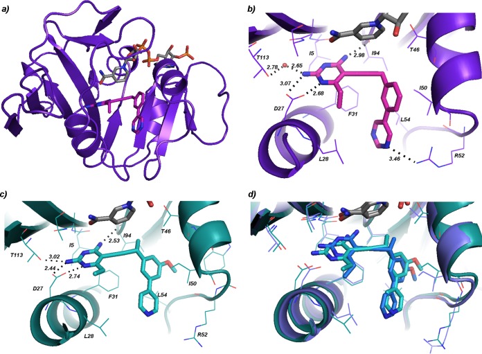
FIG 1.
K. pneumoniae DHFR bound to NADPH and propargyl-linked antifolates. (a) View of the overall structure of the sole protein chain in the KpDHFR/NADPH/compound 4 structure (NADPH in gray, compound 4 in magenta). (b) Active site of KpDHFR bound to NADPH (gray) and compound 4 (magenta). (c) Active site from chain A of the KpDHFR/NADPH/compound 3 structure (compound 3 in cyan). (d) Overlay of the active sites of chains A (cyan) and D (dark blue) in the KpDHFR/NADPH/compound 3 structure.

