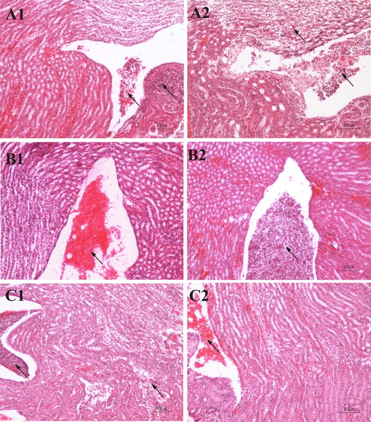
FIG 4.
Photomicrographs of H&E-stained renal sections of mice infected with ESBL-producing (A1, B1, and C1) and non-ESBL-producing (A2, B2, and C2) bacteria, at different time points, without antibiotic treatment. (A1 and A2) First day postinfection. (A1) Inflammatory lesion score 1. Neutrophils within the renal pelvic cavity (left arrow) and medulla (right arrow) are shown. (A2) Inflammatory lesion score 3. Numerous neutrophils within the pelvic cavity (right arrow) and extensive inflammatory cellular infiltration of the medullar parenchyma (left arrow) are shown. (B1 and B2) Second day postinfection. (B1) Inflammatory lesion score 1. Hemorrhage is shown with moderate numbers of neutrophils within the renal pelvic cavity (arrow). (B2) Inflammatory lesion score 2. Numerous neutrophils within the renal pelvic cavity (arrow) are shown. (C1 and C2) Sixth day postinfection. (C1) Inflammatory lesion score 3. Congestion of neutrophils within the renal pelvic cavity (left arrow) is shown. Extensive inflammatory cellular infiltration and tubular destruction in the medulla (right arrow) are shown. (C2) Inflammatory lesion score 1. Hemorrhage is shown with neutrophils within the renal pelvic cavity (arrow) and disrupted uroepithelium.

