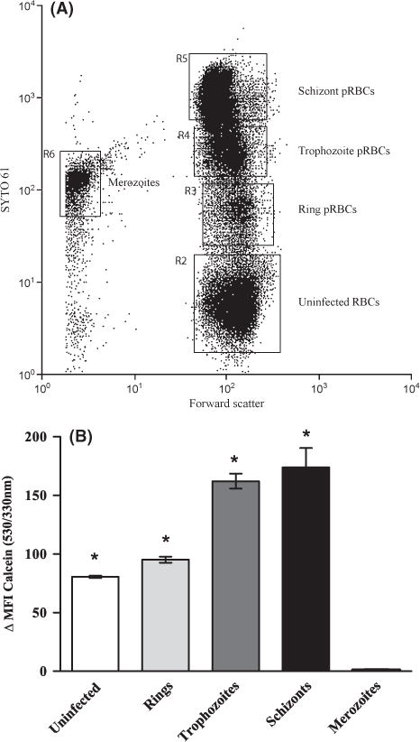
Fig 3.

The LIP increases as the parasite matures within the host red blood cell. The LIP of uninfected and different stage parasitized RBCs (pRBCs) was determined by assessing the change in MFI of calcein-loaded cells with the addition of the iron chelator, deferiprone. Cells from P. falciparum (FCR3-FMG strain)-infected erythrocyte cultures were loaded with CA-AM and then incubated with DNA dye SYTO 61 either in the presence or absence of 100 μmol/l deferiprone for 1 h and analysed by flow cytometry. (A) Dot-plot of cell distribution of DNA stain (SYTO 61) vs. forward scatter showing the distribution of uninfected RBC, ring, trophozoite, schizont pRBCs and merozoites. (B) Change in calcein MFI (530/330 nm) with the addition of deferiprone in uninfected RBCs and RBCS infected with ring, trophozoite and schizont stage P. falciparum parasites and the extracellular merozoite stage. The change in the MFI of calcein before and after addition of deferiprone represents the LIP of each cell population. The bar graph (mean ± SD, n = 3) shows calcein ΔMFI of uninfected and parasitized RBCs. Student’s t-test statistical analysis was performed comparing the MFI of calcein before and after the addition of iron chelator to cells, *P < .0002. Data is from a single representative experiment, and the experiment was performed three independent times with parasite line FCR-FMG.
