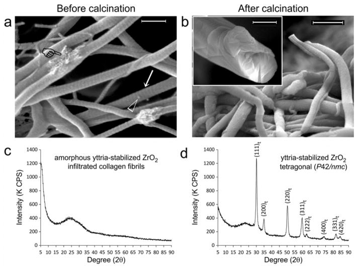
Figure 1. Infiltration of yttrium-stabilized zirconia (YSZ) into reconstituted type I collagen scaffolds.
Scanning electron microscopy images of YSZ-infiltrated collagen fibrils a) before (bar = 500 nm) and b) after calcination (bar = 500 nm). Prior to calcination, fibrils infiltrated with YSZ appear cylindrical, do not collapse when examined under high vacuum and demonstrate surface cross-banding patterns (arrow). Intrafibrillar YSZ can be vaguely discerned within fibrils that are partially squashed during handling (pointer). Open arrowhead: spherical particles probably represent extrafibrillar amorphous YSZ aggregates that have not been completely removed after rinsing the mineralized scaffolds with 1-propanol. After calcination to 800°C, cross-banding patterns disappeared in the organic matter-depleted mineralized fibrils. Inset: fractured section of a calcined fibril with a uniformly dense solid mineral core provides the evidence for successful intrafibrillar YSZ infiltration prior to calcination (bar = 200 nm). Powder X-ray diffraction of YSZ-infiltrated collagen sponges c) before and d) after calcination. Before calcination, the infiltrated mineral is probably amorphous YSZ. After calcination, the amorphous zirconia phase is partially converted into a crystalline phase, with hkl crystal lattice peak designations corresponding to tetragonal zirconia (JCPDF number 42–1164).

