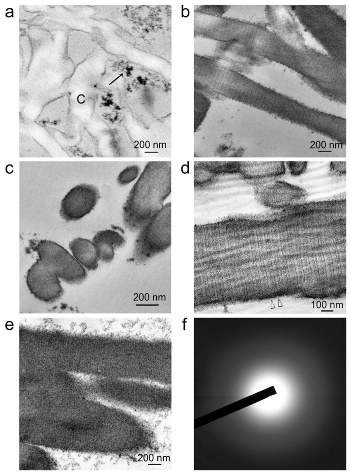
Figure 2. Transmission electron microscopy of non-osmicated, unstained sections showing the time-course of events after poly(allylamine) and glutaraldehyde pre-treated collagen scaffolds were immersed in a solution containing acac-inhibited YSZ precursors.
a) After 24 hours, electron-dense extrafibrillar YSZ precursors (arrow) can be seen around the non-infiltrated collagen fibrils (C). b) After 4 days, discontinuous electron-dense, amorphous (selected area electron diffraction not shown) intrafibrillar minerals can be discerned within some collagen fibrils. Cross-banding can be vaguely discerned in those infiltrated regions (pointers). c) Cross section of ziconified collagen fibrils showing mineral infiltration from the surface to the center of the fibrils. d) After 8 days, intrafibrillar infiltration of amorphous YSZ is intense enough for distinctive cross-banding patterns with 67 nm wide D-spacing (between open arrowheads) and rope-like microfibrillar architecture to be visible across the entire length of the collagen fibrils. e) After 12 days, collagen cross banding and microfibrillar architecture are obscured by the densely infiltrated, intrafibrillar amorphous YSZ. f) Selected area electron diffraction reveals the amorphous nature of the infiltrated minerals in e).

