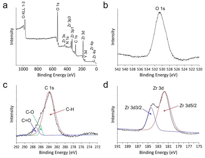
Figure 3. X-ray photoelectron spectroscopy (XPS) characterization of YSZ-infiltrated collagen scaffold.
a) Survey spectrum of the as-prepared, non-calcined sponges. b) High resolution spectrum of O 1s reveals only one peak at 530.8 eV that is derived from ZrO2. Absence of OH is indicative of complete conversion of Zr(OH)4 into amorphous zirconia. c) High resolution deconvoluted spectrum of C 1s reveals 3 peaks at 283.9, 286.0 and 287.2 eV that may be assigned to the C-H, C-O and C=O bonds, respectively. These three peaks are derived from the organic components of the collagen matrix. d) High resolution deconvoluted spectrum of Zr 3d reveals two peaks at 181.8 and 184.4 eV that may be assigned to Zr 3d5/2 and Zr 3d3/2, respectively. The detected binding energies are higher than those reported for Zr metal (180.0 eV) and ZrOx (0<x<2, 181.4 eV).

