
Figure 3.
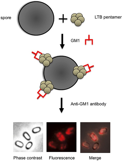
Display of a multimeric antigen on the spore surface. Purified spores are reacted with the LTB pentamers. Spore-adsorbed pentamers are reacted with the purified receptor (GM1). Spores are visualized by immunofluorescence microscopy with anti-GM1 primary antibody and Texas red conjugated secondary antibody [36]. The same microscopy field is observed by phase contrast and fluorescence microscopy. The merge of the two images is also shown.