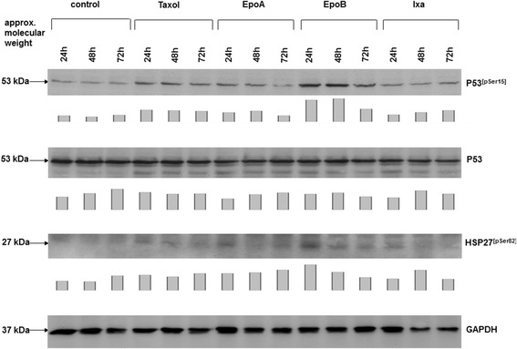
Figure 3.

Western blot analysis was performed in SCCOHT-1 cells, treated with 4 nM of either taxol, epothilone A (EpoA), epothilone B (EpoB), or ixabepilone (Ixa) for 24 h, 48 h, and 72 h, respectively. Protein aliquots of the corresponding cell homogenates were analysed for the expression of p53, phosphorylated p53 at serine-15, and phosphorylated heat shock protein (HSP27) at serine-82. The unaltered expression of GAPDH served as a control for equal loading. Quantification of the blots by densitometry scanning was normalized against the appropriate GAPDH expression and the relative expression levels were documented as bar diagram below the corresponding Western blots.
