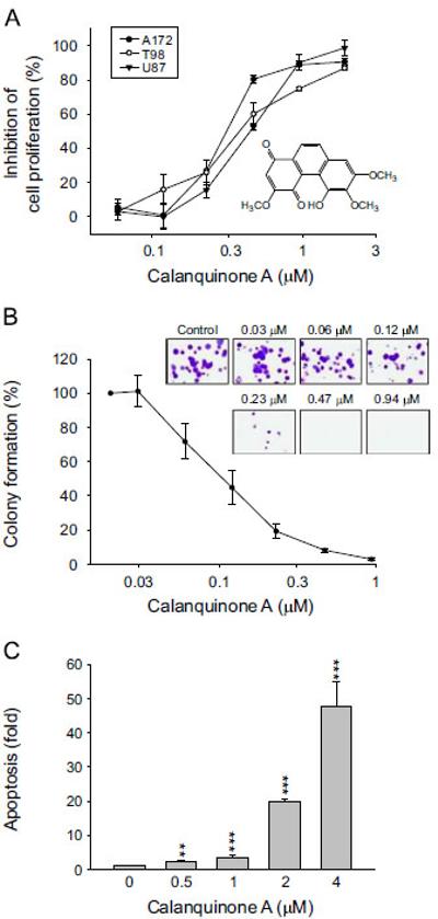
Fig. 1.

Effect of calanquinone A on cell proliferation and apoptosis of glioblastoma cells. Chemical structure of calanquinone A (A). The graded concentrations of calanquinone A were added to the cells for 48 h (A and C) or 10 days to A172 cells
(B). After the treatment, the cells were fixed and stained for SRB assay and clonogenic assay, respectively (A and B), or the DNA fragmentation was determined by the detection of nucleosomal DNA (C). Data are expressed as mean7S.E.M. of three to four determinations. nnPo0.01 and nnnPo0.001 compared with the control.
